• Home
  • Ezer Topógrafo Corneano Onyx Full

Ezer Topógrafo Corneano Onyx Full

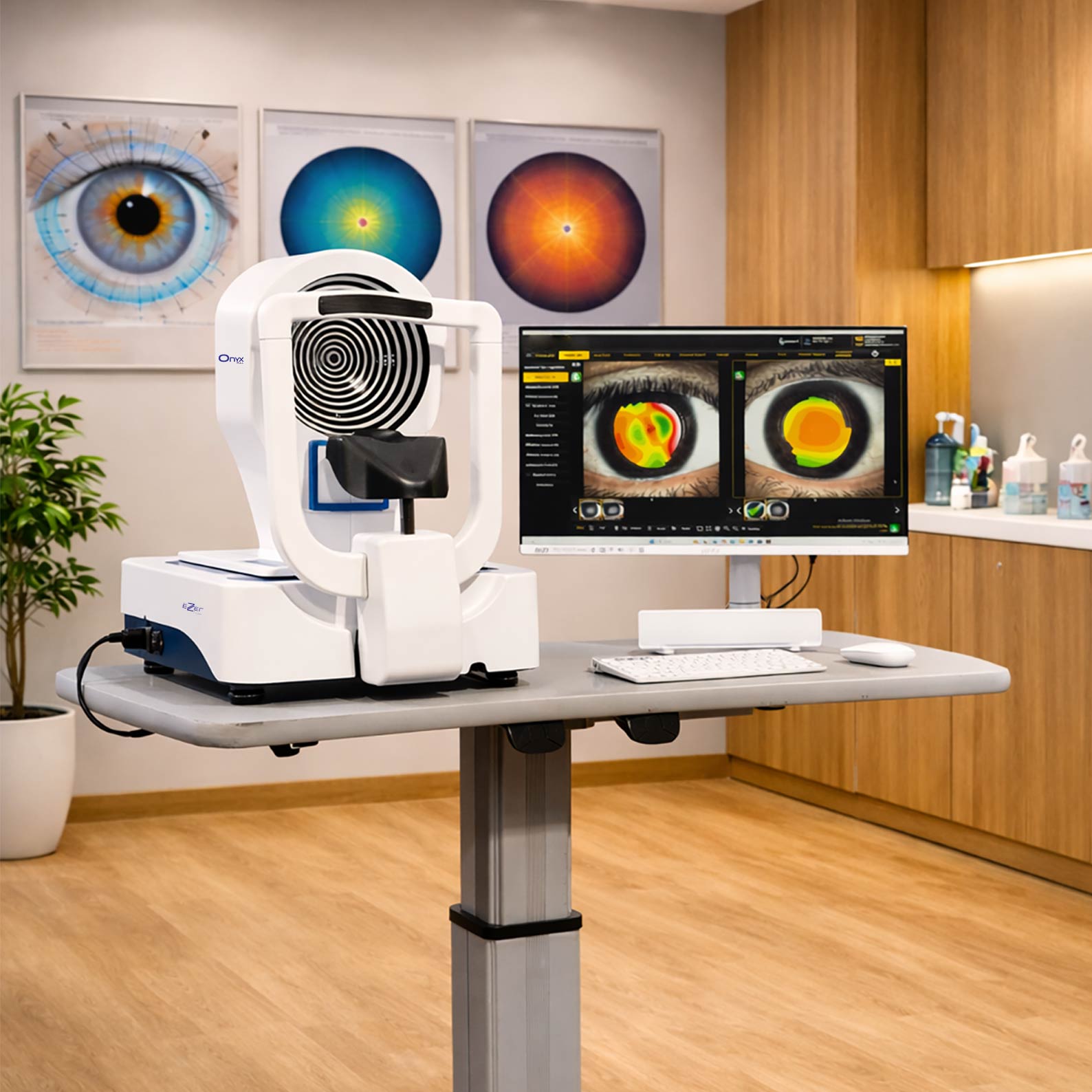
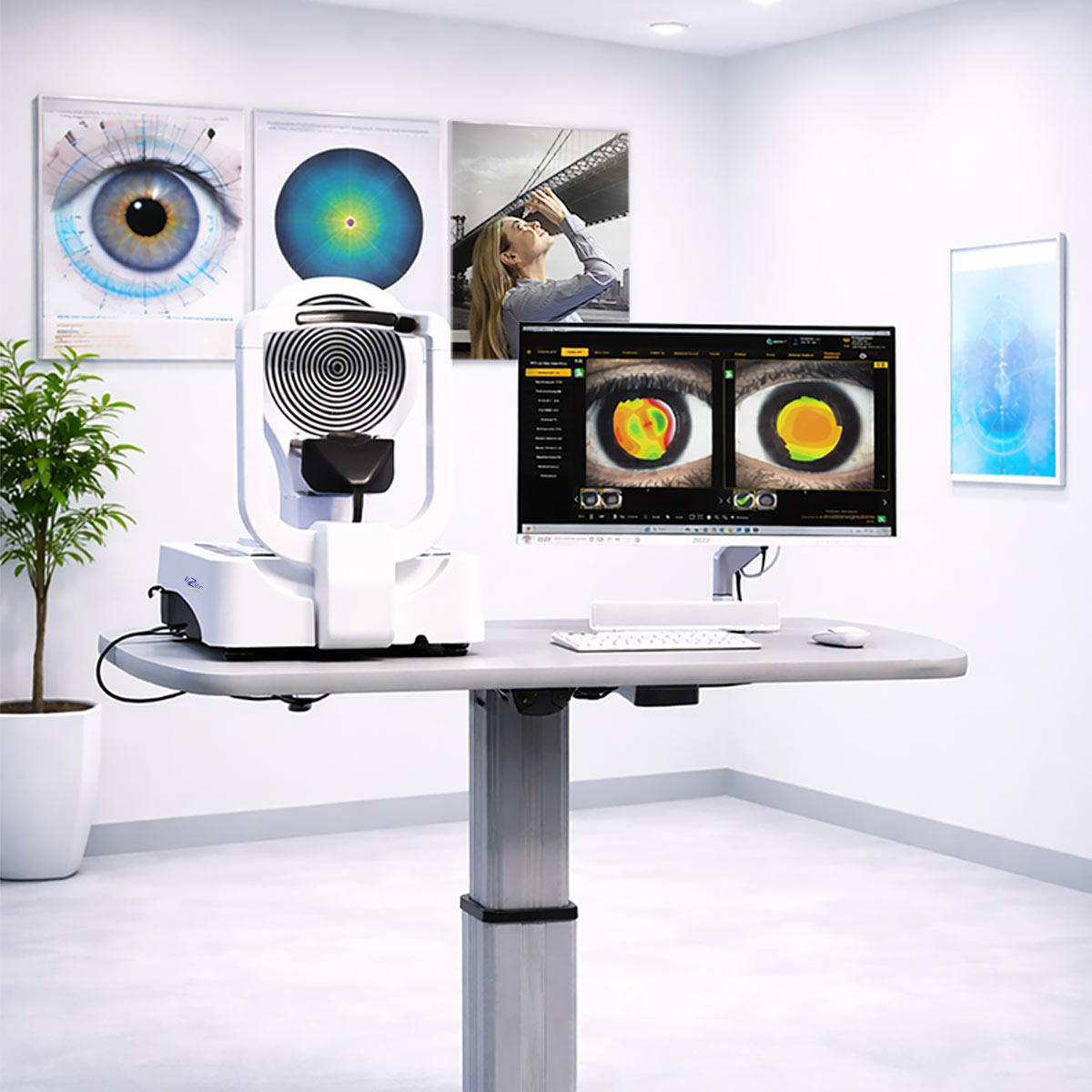
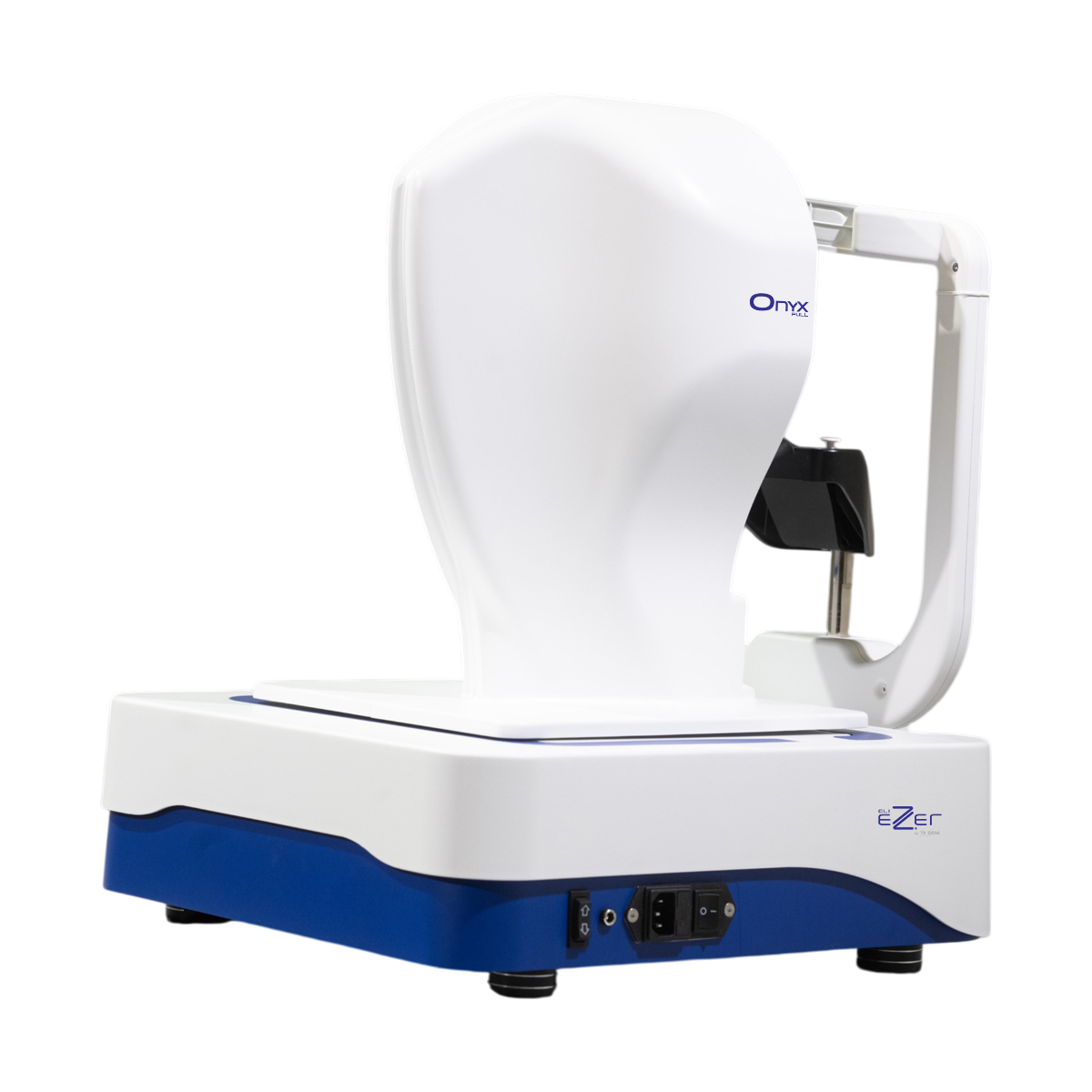
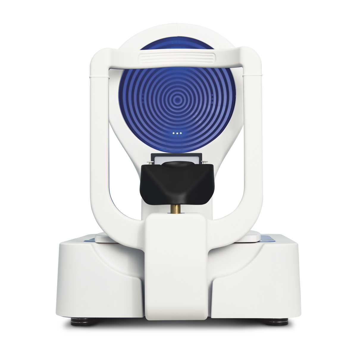
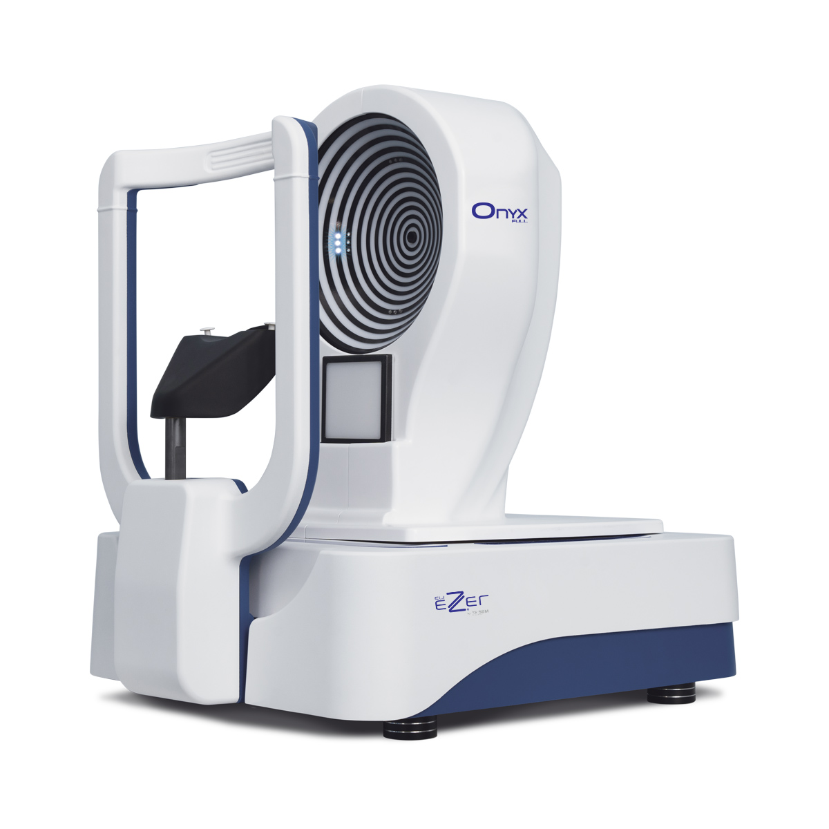
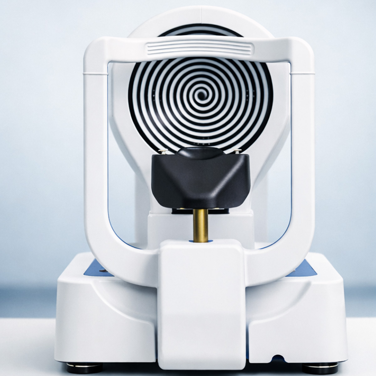
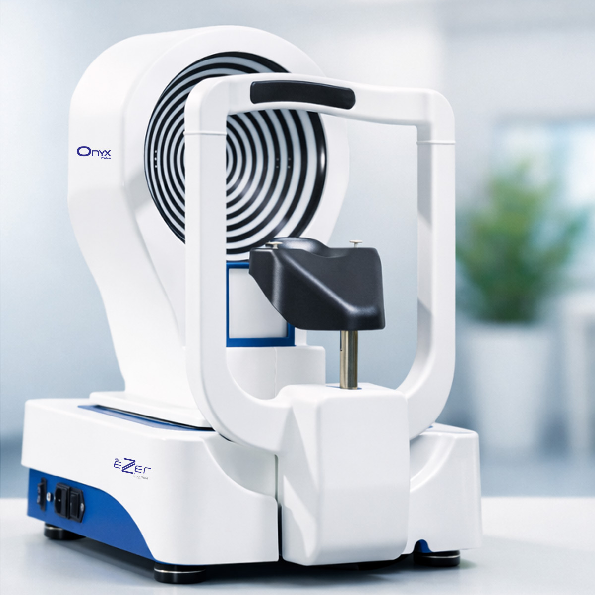
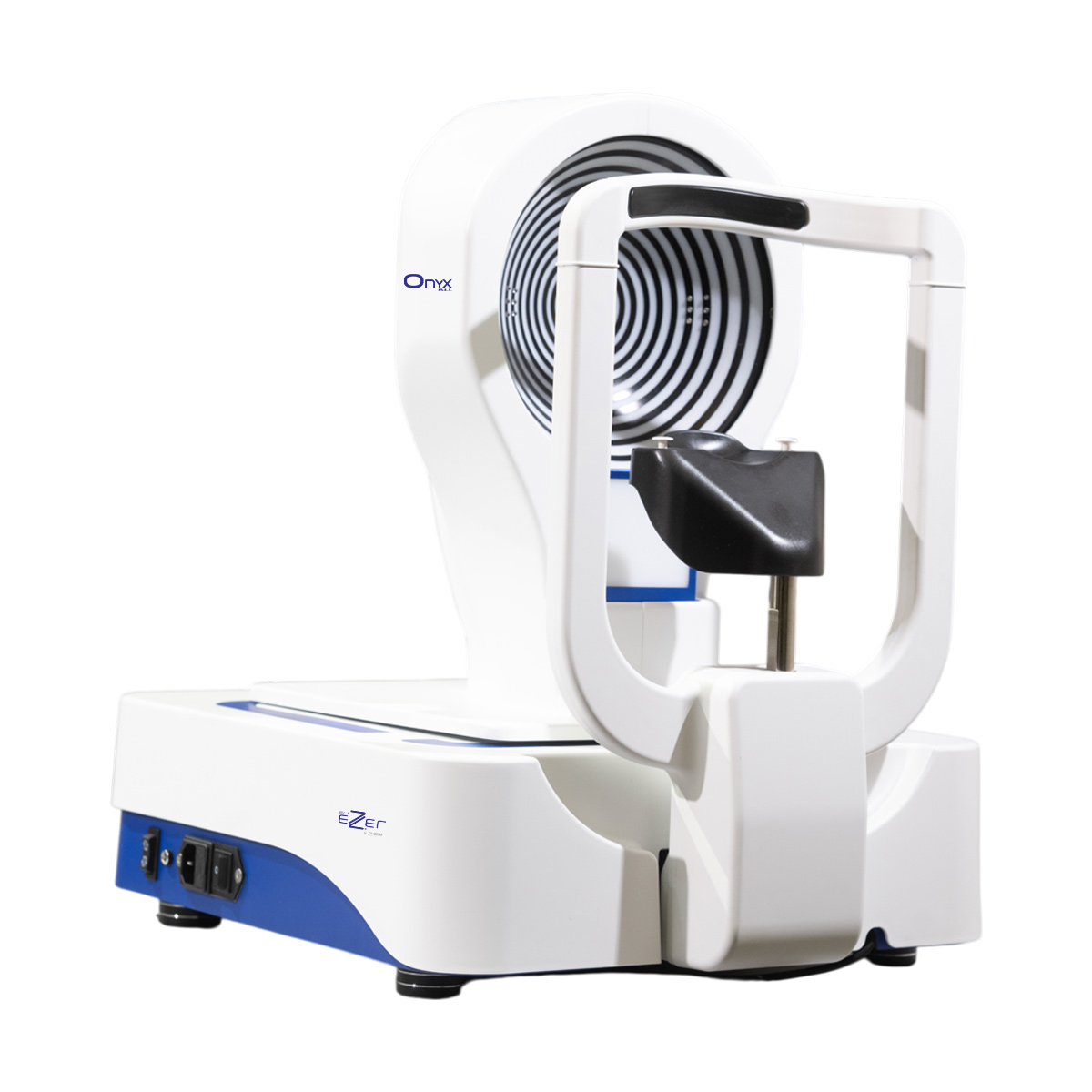
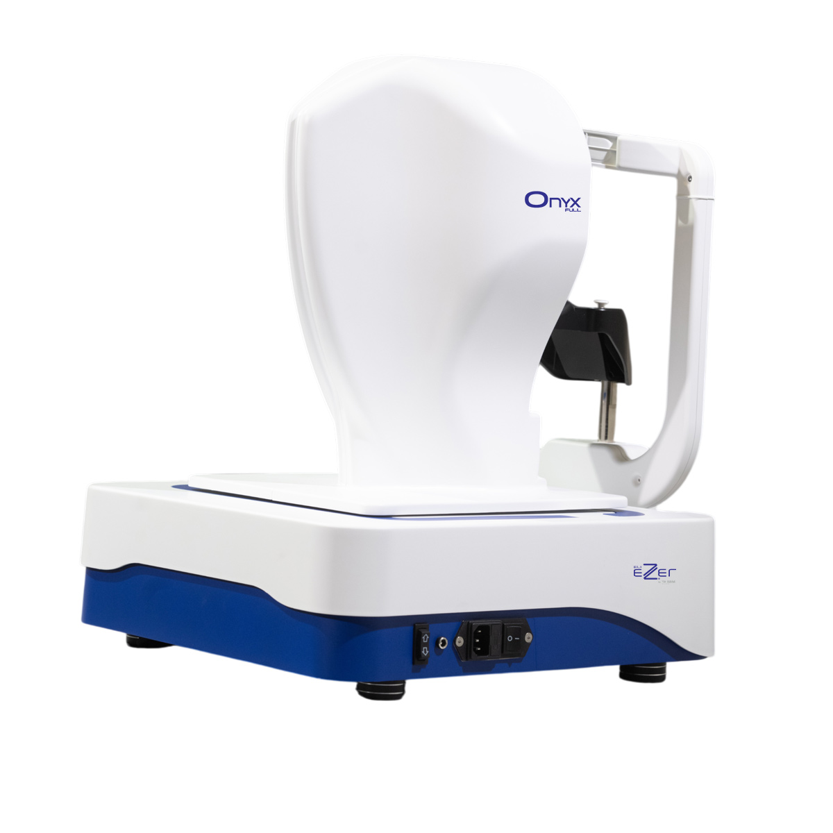
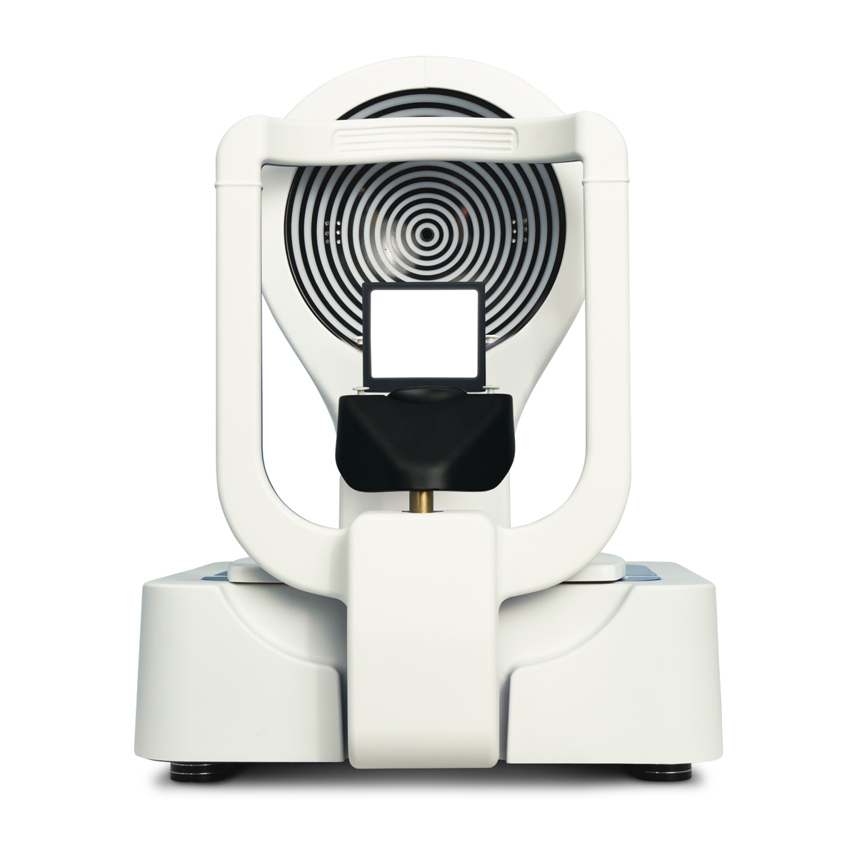
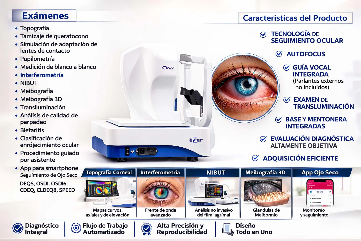
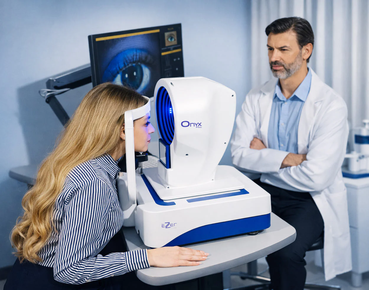
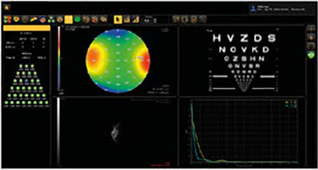
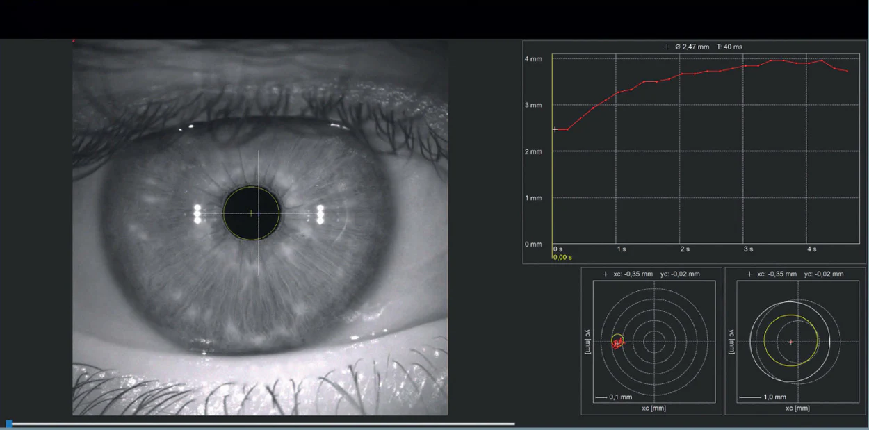
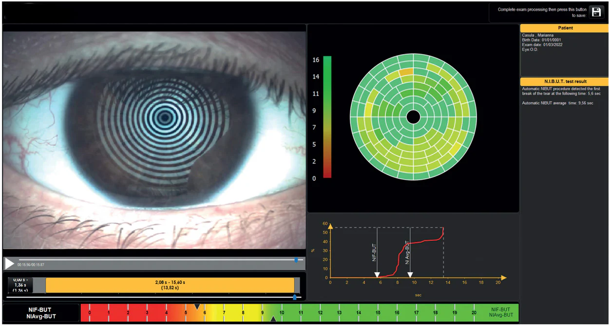
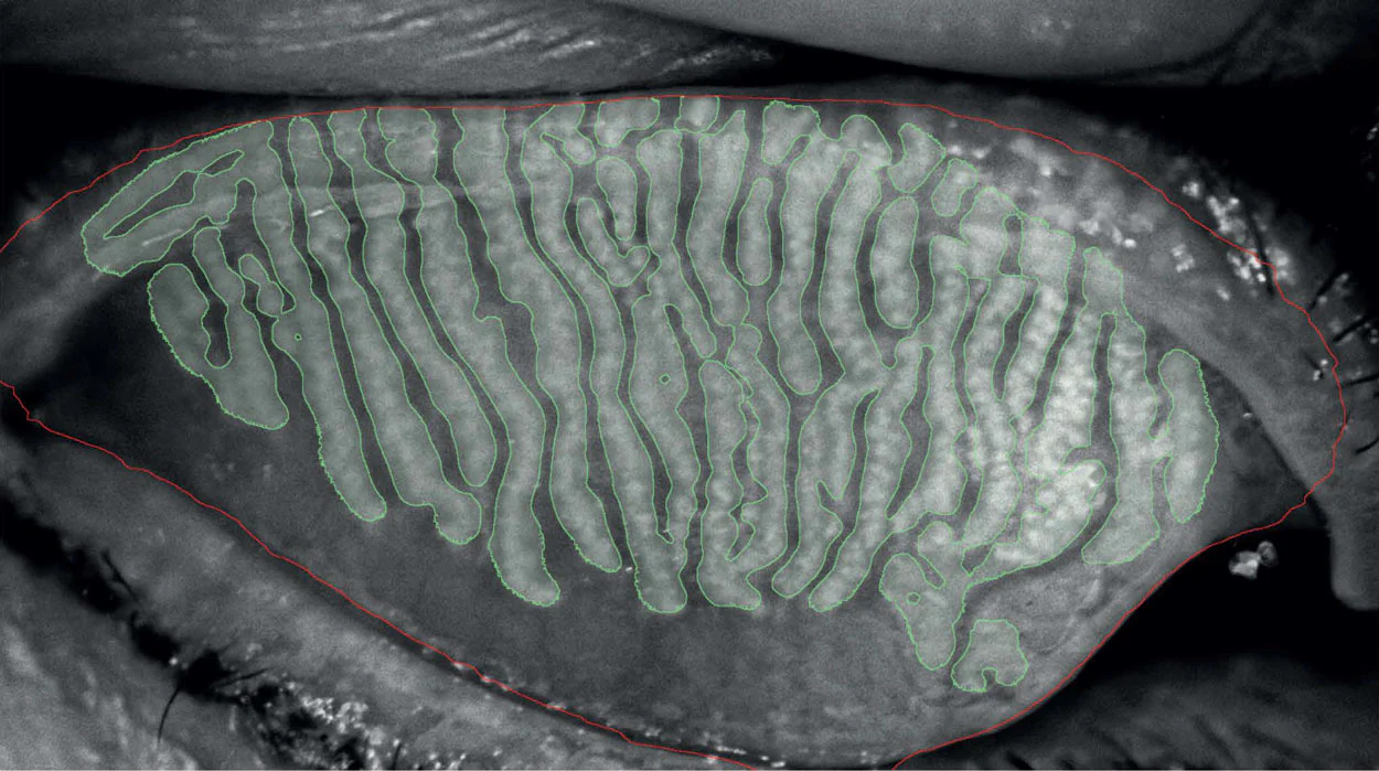
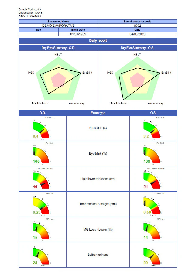
O Topógrafo Corneano Onyx Full é um dispositivo diagnóstico avançado projetado para atender às diversas necessidades dos profissionais da saúde visual. Com sua variedade de mapas especializados, como os mapas Axial, Tangencial, de Refração, Gaussiano e de Elevação, ele fornece medições corneanas precisas para avaliar a forma, a curvatura e os erros refrativos. Além disso, oferece funções inovadoras, como os Mapas Diferenciais e de Comparação, que ajudam a acompanhar as mudanças ao longo do tempo, tornando-o indispensável para monitorar condições progressivas como o ceratocone. Da mesma forma, o Onyx Full inclui ferramentas essenciais para a simulação de adaptação de lentes de contato, pupilometria, avaliação do filme lacrimal e meibografia, permitindo uma avaliação abrangente da saúde ocular. A incorporação de um aplicativo para smartphone, “Dry Eye Follow-Up”, aumenta o engajamento e o acompanhamento dos pacientes, especialmente no manejo do olho seco.

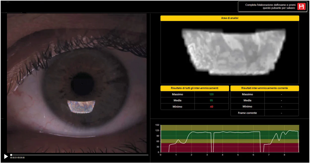
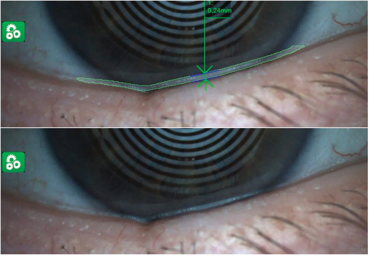
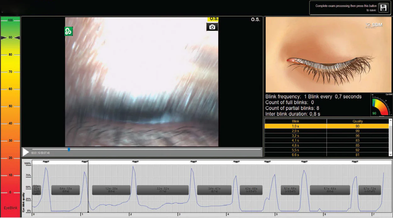
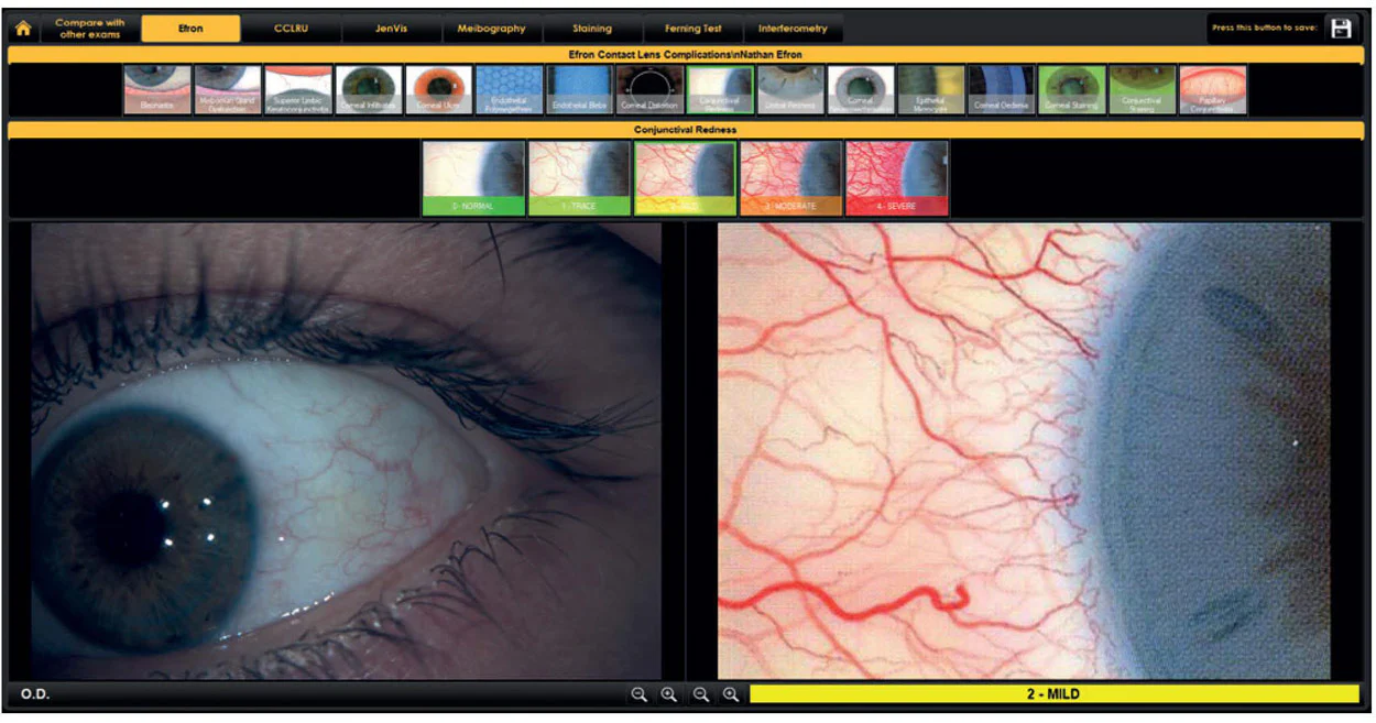
Além disso, a versatilidade do Onyx Full se estende à saúde ocular, com recursos para avaliar a hiperemia ocular, a qualidade do piscar e a blefarite. Ele também oferece suporte ao planejamento de tratamentos por meio da Seção de Protocolos de Tratamento, fornecendo recomendações personalizadas com base nos achados diagnósticos. Esse conjunto completo de funcionalidades permite que os profissionais da saúde visual ofereçam tratamentos precisos e personalizados para cada paciente, além de se comunicarem de forma eficaz com eles, tornando o Onyx Full uma ferramenta inestimável nas práticas modernas de cuidado ocular.

Falar com um especialista

The Onyx Full is an innovative diagnostic device that combines advanced corneal topography with a comprehensive dry eye assessment system, offering a complete analysis in approximately three minutes through a single scan. The equipment automatically detects the patient’s eye, adjusts for optimal focus, and initiates a complete series of ocular surface assessments, ensuring accuracy and efficiency. Its integrated system allows clinicians to objectively evaluate anterior corneal aberrations, classify dry eye subtypes (evaporative or aqueous deficiency), analyze eyelid margin anomalies, and identify inflammatory markers on the ocular surface. With a fully automated workflow that optimizes data acquisition and analysis, the Onyx Full enhances diagnostic accuracy, promotes early detection, optimizes treatment planning, and contributes to better long-term patient outcomes.

In the Onyx Full , the different light modes correspond to specific illumination technologies designed to evaluate distinct structures and parameters of the ocular surface during dry eye assessment:

Ocular surface disorders represent a significant clinical concern, making it essential to have advanced diagnostic devices that allow for accurate and comprehensive patient assessment. Among these conditions, dry eye disease is one of the most prevalent, affecting a large part of the population. Proper assessment of dry eye is especially important, as severe cases can compromise corneal health and alter its shape, influencing topography results. Modern lifestyle risk factors—such as prolonged screen exposure, air conditioning use, and environmental pollution—are strongly related to the development and chronic progression of dry eye, potentially impacting other ocular conditions over time. Implementing an automated, structured, and personalized management strategy can significantly improve therapeutic outcomes and patient quality of life. Furthermore, an AI-based algorithm enhances the captured image to up to 23 megapixels , generating ultra-high-quality images that allow for a new level of precision in Placido disk-based corneal topography measurements.

Onyx Full – Key Advantages for Workflow Innovation

🔹 Ease of Use
Save time and reduce staff workload with a system that manages focus and acquisition fully automatically. Equipped with an innovative automatic mechanism, our Placido disc-based corneal topographer autonomously detects and focuses on the patient’s eye, eliminating the need for manual adjustments by the operator and simplifying the examination process.

🔹 High Precision
Automated functionality improves the accuracy of both topographic measurements and dry eye assessment, ensuring greater diagnostic accuracy. Advanced autofocus technology increases the reliability and repeatability of corneal measurements. Each examination is optimized to deliver fast, consistent, and highly accurate results, making Onyx an essential tool for any ophthalmology clinic or practice.

🔹 Operator and Patient Comfort
Reduced manual intervention minimizes the risk of operator-dependent errors and improves patient comfort throughout the examination. An optimized workflow creates a more agile experience for both clinicians and patients.

Key Features and Workflow
Equipped with an automated base, the Onyx Full initiates the data acquisition process with a single click. Once activated, the device automatically aligns with the patient’s eye, adjusts focus, and captures the necessary images, all guided by an intuitive software interface. A customized examination protocol then directs the workflow, automatically running selected diagnostic tests and processing results with minimal user intervention.

Unique Technology for Automated and Objective Analysis of Patients with MGD (Meibomian Gland Dysfunction)
Tear film interferometry is increasingly used in research to observe the tear film. Interferometry is a technique that examines the surface refractive patterns and dynamics of the lipid layer, allowing the measurement of tear film stability and lipid layer thickness.

Interferometers are applied research tools in many fields of science and engineering. They are called interferometers because they work by combining two or more light sources to create an interference pattern that can then be measured and analyzed.

The resulting patterns can appear as meshes and waves, just waves, or colored fringes. Interferometry specifically studies the lipid content of the tear film. The lipid layer, the outermost component of the tear film, protects the underlying aqueous layer from evaporation. Lipid layer deficiency is common in many patients with MGD, making this assessment especially important.

The Onyx Full system features advanced, non-invasive corneal topography that generates a precise map of corneal surface curvature. This essential diagnostic tool allows for early detection and monitoring of keratoconus, improves pre- and post-operative assessment in refractive surgery, and ensures precise contact lens fitting.
By capturing detailed curvature measurements, the system produces an intuitive, color-coded map, where each color represents a different corneal power. This allows professionals to quickly identify irregularities, assess the impact of contact lenses on the cornea, and optimize lens selection to achieve maximum patient comfort and visual performance.

Aberrometry Analysis (Zernike)
The ONYX Corneal Topographer performs aberrometry analysis using Zernike polynomials. This analysis provides data on astigmatism, spherical aberrations, higher-order aberrations and coma, as well as Optical Path Difference (OPD). It covers pupil sizes ranging from 2.5 mm to 7.0 mm, offering a comprehensive view of corneal characteristics.

Advanced Altimetry
: Advanced altimetry involves creating a differential map that compares a reference surface with the patient’s eye. This map is decomposed into Zernike polynomials, specifically up to the seventh order, offering a detailed analysis of the eye’s topographic characteristics.

Visual Acuity Assessment
: Visual acuity assessment involves simulating the patient’s visual clarity considering Zernike wavefront aberrations. This simulation helps illustrate the impact of conditions such as cataracts and the results of refractive surgery on the patient’s vision.

Comparison of Exams / Differential Map
The “comparison of exams” visualization graphically presents the changes that occur over a specific period, such as the progressive development of conditions like keratoconus. This function facilitates the explanation of even complex scenarios to your patients, allowing for clear communication.
It is possible to compare up to four exams simultaneously, expanding your ability to illustrate changes and trends.
Furthermore, the “differential map” visualization highlights the differences between two selected maps, with the option to use up to three exams for this purpose.

The auto-fit module combines topographic data with RGP lens data to determine and customize the best solution for the patient’s eye, simulating the fitting process with fluorescein. With Onyx Full , it is possible to capture in vivo fluorescein images of the lens or assess the fit through simulated fluorescein visualization.
Contact lens simulation generates an image illustrating how a specific lens fits the eye. This simulation allows for adjustment of the angle and position of the contact lens, with automatic recalculation of the fluorescent image. The system streamlines the ordering process, reducing the number of lenses needed and minimizing consultation time, while improving the first fitting success rate. This process simplification is achieved by providing real tear film-independent elevation data.

With the ONYX Full , you can efficiently and accurately collect all the essential data needed for multifocal, bifocal, and toric contact lenses.
Pupillary measurement involves capturing images or videos in both white light and the infrared spectrum. It allows for precise measurement of pupil size and decentration under various lighting conditions, including scotopic, mesopic, and photopic vision.
This method offers a practical and quick way to assess your patients’ pupil size in different lighting scenarios. This functionality not only assists in the fitting of multifocal lenses but is also extremely valuable for measuring the optical zone before refractive or cataract surgery.

Dynamic Pupilometry:
Measuring pupil diameter has gained increasing importance in the field of refractive surgery. Larger scotopic pupil sizes can influence the development of postoperative symptoms such as halos, glare, and monocular diplopia. Accurate pupil diameter measurements under scotopic conditions are essential for refractive surgeons, as they help determine appropriate treatment zones in excimer laser procedures, corneal treatments, and intraocular surgery.
Dynamic pupillometry is a method that observes the transition from a well-lit photopic environment to a low-light scotopic environment. This technique functions as a simple screening tool to quantify the pupillary light reflex (PLR), providing information about the activity of the autonomic nervous system (ANS). Infrared videography is used to measure the pupillary light reflex, which is subsequently classified into several quantitative parameters that act as indicators of ANS activity.

The ONYX Full is recognized as the fastest and most comprehensive dry eye system available. The Tear Film and Ocular Surface Society study clearly defined the critical parameters for diagnosing dry eye syndrome through a comprehensive analysis of the ocular surface. A fundamental initial step in this screening process is classifying the pathology subtype through a detailed examination of both the lipid and aqueous layers of the tear film.
The ONYX Full excels at facilitating this comprehensive analysis, applying established guidelines in a fully automated and objective manner. It generates results automatically, eliminating the need for operator intervention, making the analysis extremely fast and easily delegable to other team members. This innovative approach significantly improves the efficiency and accuracy of dry eye diagnosis.

NIBUT Automatic

The assessment of mucin layer and tear film stability in general is performed using non-invasive breakup time (NIBUT) analysis, utilizing Placido rings projected onto the cornea. This test allows for the automatic evaluation of tear film stability without the need for fluorescein staining.
The main parameters automatically evaluated during NIBUT analysis include:
First NIBUT
NIBUT Medium
NIBUT Map
Tear Film Dynamic Graph (TF Dynamic Graph)

Interferometry

According to TFOS DEWS II, tear film instability and lipid layer dysfunction are central mechanisms of dry eye disease, with the evaporative form being the most prevalent worldwide; therefore, evaluating the lipid layer is no longer optional, but essential. Automated interferometry provides a direct and objective assessment of this layer, allowing clinicians to identify the root cause of symptoms and make more precise therapeutic decisions, especially considering that most treatments target evaporation and Meibomian gland dysfunction. With automated focus, acquisition, and analysis, the examination becomes reproducible, operator-independent, and suitable for follow-up, making interferometry a reliable and evidence-based diagnostic tool.

Automatic Height of the Tear Meniscus

Insufficient tear production can cause symptoms of dry eye, commonly referred to as aqueous deficiency (AD). However, accurately measuring tear volume can be complex, as many available methods are invasive and cause discomfort to patients.
An alternative approach to assessing tear volume involves examining the thickness of the tear meniscus observed along the eyelid margins. This meniscus can be evaluated by considering its height, regularity, and shape, providing valuable information about tear volume and tear film stability.
To optimize this process, artificial intelligence plays a key role by automatically determining essential parameters such as the position of the tear meniscus and identifying the maximum value within it. This automation not only improves the accuracy of the assessment but also minimizes patient discomfort during the examination.

Lisamin Green: Conjunctival and Palpebral Staining

In the management of dry eye, assessment of the ocular surface should not be limited to the cornea alone. The conjunctiva and eyelid margin often show the first signs of cellular stress. Lisamine green staining highlights stressed, damaged, or devitalized cells in these areas, providing a more complete assessment of ocular surface health. This is especially valuable in chronic and evaporative dry eye, where conjunctival involvement may appear before—or be more evident than—corneal findings.
The Onyx Full allows for the acquisition of high-quality images with Lisamine green, facilitating clear documentation and reliable comparisons over time for accurate follow-up.

Infrared Imaging:
Meibomian glands produce the lipid layer of the tear film, and their dysfunction is strongly associated with evaporative dry eye. Assessing both their structure and function is therefore essential to understanding the origin of the condition. Meibography allows for a clear visualization of the glandular architecture, offering information on remaining secretory capacity and potential for functional recovery. The Onyx Full system provides a detailed and easy-to-interpret visualization that can also be easily understood by the patient.

Eyelid Transillumination:
What makes Onyx Full different?
Its infrared transillumination function reveals critical details that often go unnoticed during routine examination, allowing visualization of glandular dysfunction, lipid accumulation, and vascular changes in the eyelids for a more complete assessment of dry eye

Meibomian Gland 3D:
Incorporating three-dimensional images of the Meibomian glands offers two main advantages. First, it allows for direct comparison with a healthy eye, facilitating the clear identification of abnormal glandular structures through three-dimensional visualization. Second, it generates intuitive images that can be shared with patients, helping them to better understand the underlying cause of their discomfort.

Blinking Quality: Glandular Stimulation

Blinking is essential for evenly distributing the tear film and promoting the expression of the Meibomian glands. Incomplete or reduced blinking can compromise tear film stability and worsen dry eye symptoms. Onyx Full automatically analyzes blink dynamics, providing valuable information about the functional role of the eyelids in the disease.

Blepharitis: Inflammation of the eyelid margin

Eyelid margin disorders are a frequent cause of tear film instability and ocular discomfort. Findings such as cylindrical dandruff and other signs of blepharitis indicate chronic inflammation that can also affect the function of the Meibomian glands. Onyx Full allows for clear documentation of the eyelid margin, facilitating accurate diagnosis and monitoring the effectiveness of targeted treatments.

Demodex: Source of Eye Irritation

Demodex is a microscopic mite that can colonize the eyelid margin and is associated with inflammation, eyelash changes, and persistent irritation when the infestation is significant. Its evaluation is especially important in cases of recurrent or treatment-resistant blepharitis. Onyx Full allows visualization and documentation of related clinical signs, facilitating a more complete and specific diagnosis.

Staining: Documentation of Epithelial Damage

Staining the ocular surface is crucial for assessing epithelial integrity, as changes in the tear film and environmental stress can cause cellular damage. By highlighting affected areas of the corneal and conjunctival epithelium, staining helps differentiate dry eye from other pathologies with similar symptoms and determine its severity. The Onyx Full captures and stores staining images, allowing for reliable monitoring and treatment control over time.

Bulbar Hyperemia: Inflammation of the Ocular Surface

Bulbar hyperemia is a key indicator of ocular surface inflammation and generally reflects ongoing inflammatory processes. Its presence, distribution, and intensity provide valuable information about conjunctival health and disease progression. The Onyx Full records standardized images of the bulbar conjunctiva, facilitating consistent assessment and long-term follow-up.

Comparative Tables (Efron / CCLRU / Jenvis): Clear Clinical Communication

Standardized grading scales such as Efron, CCLRU, and Jenvis allow professionals to compare clinical findings with established references, improving diagnostic clarity and communication. Onyx Full integrates captured images with these reference tables, incorporating the classification directly into the clinical report for objective documentation and accurate follow-up.

Multiple Reports Available
The software functions as a specialized platform for the management of dry eye. In addition to assisting in the diagnosis and classification of the disease, it allows for the generation, printing, and storage of various medical reports, ensuring professional and clinically sound solutions for patients. Providing technical documentation related to the examinations performed is a common practice that increases patient satisfaction. The different print formats available through the SBM device allow professionals to visually explain the pathological situation and effectively communicate the progression of the disease, facilitating the development of personalized treatment plans.

Daily Report:
The “daily report” consolidates all the results of the tests performed on the patient on a specific date. It offers multiple viewing options, allowing the information to be presented with or without images, adapting to the specific needs of both the healthcare professional and the patient.

Follow-up Report
The “follow-up report” is a valuable tool for demonstrating the benefits of therapy to the patient. It visually shows how the treatment has improved dry eye symptoms, providing a clear and tangible representation of the results achieved.

The ” All
Reports” function allows you to generate a complete dossier of up to 16 pages with detailed information. This is especially valuable when the patient bears the cost of the service, as it adds transparency, comprehensiveness, and a tangible representation of the care received.

Treatment Report
The “treatment report” presents a complete list of all therapies prescribed to the patient, offering a clear and organized record for consultation and follow-up.

This is a smartphone application developed to offer patients access to their test results, information about their therapy, and comprehensive knowledge about dry eye disease.
The platform provides interactive, fast, and convenient communication between doctor and patient, establishing an efficient two-way connection. It allows for the accurate sharing of reports and updates, helping patients track their progress over time. Furthermore, it facilitates the regular scheduling of appointments and interaction with the clinic or hospital. The system also automates the sending of reminders to patients and notifications to professionals, optimizing appointment management and the implementation of prescribed treatments.